Hydnophlebia chrysorhiza
Scientific name: Hydnophlebia chrysorhiza (Eaton)
Parmasto
Derivation of name: Chrys- means "golden" or "gold" and
rhiz- means root." Chrysorhiza means "golden root" in
reference to the bright yellow rhizomorphs.
Synonyms: Phanerochaete chrysorhiza (Torr.)
Budington & Gilb.; Hydnum chryscomum;
Mycoacia fragilissima; Oxydontia fragilissima.
Common name(s): Spreading yellow tooth.
Phylum: Basidiomycota
Order: Polyporales
Family: Meruliaceae
Occurrence on wood substrate: Saprobic; sheetlike and
flattened against the underside of hardwood logs; June through
January.
Dimensions: Fruit bodies 2-6 cm wide or much larger;
cordlike, bright orange, branching rhizomorphs may be 10-20
cm long.
Description: This fungus appears as a bright orange-yellow
spreading crust with a white margin and with bright orange
rhizomorphs serving as runners. Close examination reveals the
fertile surface is made up of crowded, yellow to orange
spines with rounded tips.
Comments: Finding this colorful fungus is worth the effort of
looking at the underside of logs.
More information at TomVolkFungi.net:
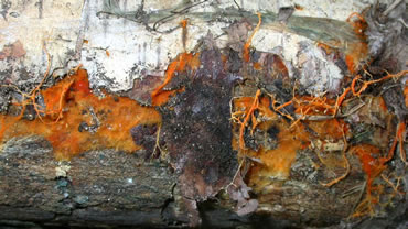
Figure 1. This fungus was observed on the undersurface of a
log in Wisconsin. Although the spiny fertile portion was not
present, Tom Volk and I concluded that the presence of the
bright orange rhizomorphs was good evidence this specimen
is Hydnophlebia chrysorhiza. Photo © Gary Emberger.
%20Brookhaven%20NY%20Tom%20Bigelow.jpg)
Figure 2. This award-winning photograph of spreading
yellow tooth shows spines in the center, a white growing
margin, and orange rhizomorphs.
Photo © Tom Bigelow.
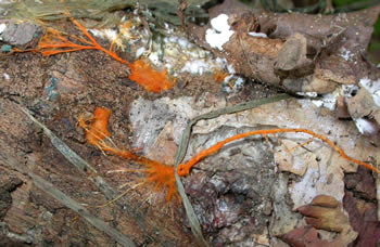
Figure 3. The rhizomorphs function to enter the wood
substrate. Photo © Gary Emberger.
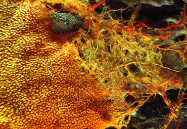
Figure 4. The fertile, spine-bearing portion of
Hydnophlebia chrysorhiza is to the left and the sterile
rhizomorphs are at the right. Photo © Dianna Smith.
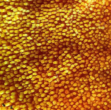
Figure 5. Enlargement of a portion of Figure 4 showing the
rounded spines. Photo © Dianna Smith.